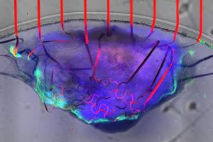

Artists impression over photo:
“Cardiac tissue is very special,” said Jun Yao of UMass Amherst’s college of engineering. “It has a mechanical activity – contractions and relaxations that pump blood through our body – coupled to an electrical signal that controls that activity.”
‘Cardiac micro-tissue’ is the material of interest, according to the university, which is grown from human stem cells and is vulnerable to damage if sensors are inserted after growth. Hence the need to grow on a pre-existing matrix of sensors, in a way that allows it to grow unimpeded.
Monitoring ‘excitation-contraction coupling’ in the sample was the aim of the experiment, by measuring both the self-generated electrical stimulus from the tissue and any resulting movement.
The team designed 20 x 20μm graphene transistors – roughly the size of a cell – to do the sensing, with their channels modulated by local electrical potentials through the field effect, while simultaneously detecting mechanical movement because they are naturally piezo-resistive.
“Because graphene is impossibly thin, it can register even the tiniest flutter of muscle contraction or relaxation, and can do so without impeding the heart’s function,” according to the university.
These transistors are inter-connected by a mesh of gold palladium conductors that snake their way across a flexible substrate – repurposed SU-8 photo-resist. A layer of silicon nitride over the conductors insulates them from the tissue sample.
The serpentine nature of the conductors adds not only flexibility but stretchability, resulting in a “soft, stretchable porous mesh scaffold that has close structural and mechanical properties to human tissue and which can be applied non-invasively to cardiac tissue” said UMass Amherst.
“No one has ever done this before,” according to fellow researcher Hongyan Gao. “Graphene can survive in a biological environment without degrading for a very long time and not lose its conductivity, so we can monitor the cardiac micro-tissue across its entire maturation process.”
The university worked with MIT on this project, which is described in the Nature Communications paper ‘Graphene-integrated mesh electronics with converged multifunctionality for tracking multimodal excitation-contraction dynamics in cardiac microtissues‘.